T H E O R Y
Oxygen
Free Radicals and Mitochondrial Mutation
The control region is relatively tolerant of a high mutation rate, because binding sites for DNA and RNA polymerase are defined by only short nucleotide sequences. The high mutation rate of mt DNA is almost certainly due to the fact that the mt genome is located in close proximity to the respiratory machinery of the cell - a known source of potent mutagens called oxygen free radicals.
Oxygen free radicals are a natural byproduct of respiration. Electrons formed during the oxidation of glucose are passed along the electron transport chain, a series of electron-accepting molecules embedded in the mt membrane. Protons created during electron transfer ultimately are used to drive the synthesis of ATP. In the final step of transfer, electrons are combined with oxygen and protons to produce water.
However, faulty electron transfer at any point in the electron transport chain, results in an electron being accepted by atomic oxygen(O2). The superoxide free radical created (O2. -) has a single unpaired electron (designated by the "dot" in the chemical formula), which seeks to react with an electron source to make a stable electron pair. Under physiological conditions, electrons "leak" from the electron transport chain, converting about 1-3% of oxygen molecules into superoxide.
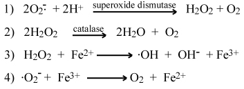
The cell has evolved a two-step mechanism to disable oxygen free radicals. In the first step, superoxide free radical is simultaneously reduced and oxidized (dismutated) to form hydrogen peroxide and oxygen (reaction 1 below). This is accomplished by superoxide dismutase, a so-called metabolic enzyme. Although hydrogen peroxide is also a reactive oxygen species, it is much less reactive than superoxide. In the second step, hydrogen peroxide is converted into water and oxygen by catalase enzymes (reaction 2). Hydrogen peroxide readily diffuses out of the mitochondria, and its level in the cytoplasm may provide the cell a means to monitor the efficiency of respiration.
Superoxide and hydrogen peroxide are not thought to be the major causes of mutations in the mt genome. Ironically, the most mutagenic of the reactive oxygen species, hydroxyl (.OH) free radical, is generated as a consequence of disabling superoxide to hydrogen peroxide. Termed Fenton chemistry, peroxide readily reacts with ferrous iron (Fe2+) or other transition metal ions to produce hydroxyl radical (reaction 3). By the same token, ferric iron (Fe3+) can accept an electron from superoxide, cycling it back to the ferrous state and making it available to react with another peroxide molecule (reaction 4). Thus, even trace amounts of iron ion can potentially catalyze the formation of large amounts of hydroxyl free radical.
Hydroxyl free radical, which is also produced when ionizing radiation strikes water, is several orders of magnitude more reactive than superoxide. The hydroxyl radical reacts immediately upon contact with a suitable electron donor, so its reaction rate essentially is controlled by the rate at which it diffuses. Hydroxyl radicals react with all types of biologically important molecules - nucleic acids, proteins, sugars, and lipids - producing radicals that undergo further reactions. DNA radicals can react with protein radicals (in histones) to form crosslinks that interfere with chromatin unfolding, DNA repair, replication, and transcription.
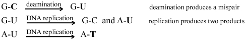
It is believed that iron ions complex with negatively charged phosphates in the DNA backbone, thus generating hydroxyl radicals within diffusion distance of reactive bonds in deoxyribose and nitrogen bases. Hydroxyl free radicals attack all positions of the deoxyribose sugar, leading to single- and double-stranded breaks in DNA. Hydroxyl radicals also deaminate nucleotides, leading to point mutations or SNPs - notably C>T, G>C and G>T changes. The C>T change is termed a transition, because C and T are both pyrimidine nucleotides. The G>C and G>T changes are termed transversions, because a purine nucleotide (G) is converted into a pyrimidine (C or T). The following shows the sequence of events that produce a C>T transition.
DNA damage by oxygen free radicals suggests an accelerating degradation of mt function over time. Accumulating mutations in the genes encoding electron transporters (NADH dehydrogenase, cytochromes, and coenzyme Q) lead to decreased transfer efficiency, which, in turn, leads to higher production of superoxide and hydroxyl free radicals. Mutations in ribosomal and transfer RNAs lead to inefficient or errant translation of proteins encoded by the mt genome. There is growing evidence for this "mitochondrial theory of aging." Since 1988, mutations in mt genes have been implicated in a number of degenerative diseases - including Alzheimer disease, mitochondrial myopathy, Kearns-Sayre syndrome, CPEO (chronic progressive external phthalmoplegia), Leigh syndrome, Pearson syndrome, dystonia, and diabetes. Not surprisingly, most of these diseases affect organs and tissues that have a high demand for energy. Mt mutations also accumulate in tumor cells, and are so highly amplified that often they can be detected in bodily fluids.
Noncommercial, educational use only.